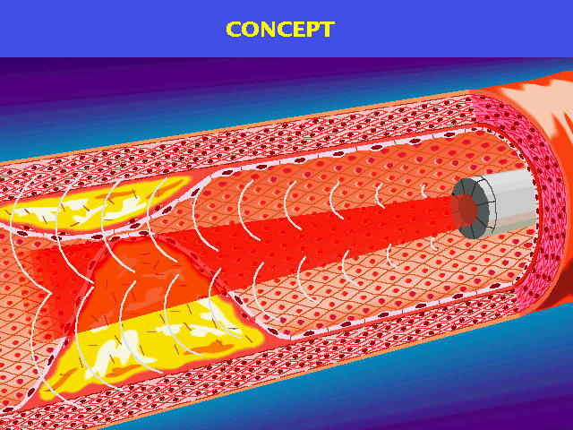
In conventional ultrasonic imaging methods,  it is impossible to image
a three-dimensional domain area instantaneously, because they requires
scanning  of a narrow and directional beam.  To overcome this problem,
we suggest  a new ultrasonic imaging method  using a ring array probe.
This method enables to obtain three-dimensional images instantaneously
using spherical pulsed waves which can illuminate a measurement domain
all at once. Hence, it does not need beam scannings.

We suppose  that  our new method  will be available  for intravascular
endoscopes. The following is a conceptual image of this research.
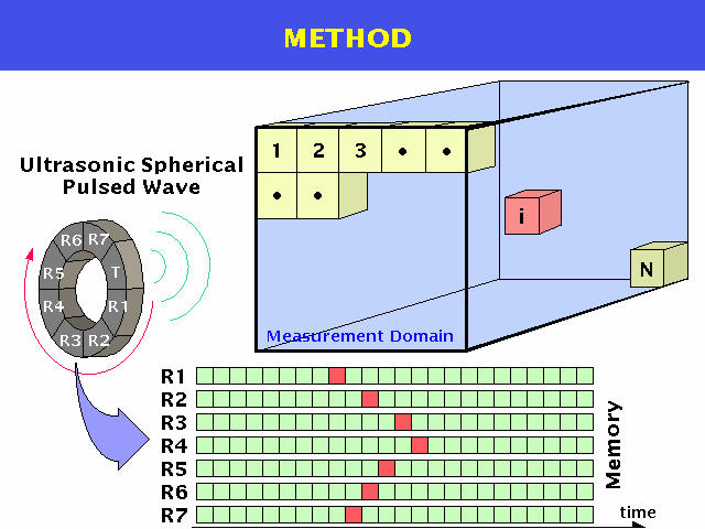
In our method,  we use a ring array probe  wich has eight transducers.
Three-dimensional frontal image  can be obtained instantaneously  with
the following algorithm.

(1)  The measurement domain is determined and is quantized into
     N voxels
(2)  An ultrasonic spherical pulsed wave is transmitted  from a
     transducer to the measurement domain
(3)  Echo signals  from the measurement domain are received  by
     the other seven transducers  and are stored in each recei-
     ver's memory  after A/D conversion and filtering
(4)  The propagation time  of the pulsed wave is calculated for
     each receiver (transmitter - voxel No.i - receiver)
(5)  The absolute values of data  corresponding to the propaga-
     tion time are taken from each receiver's memory,  and they
     are summed
(6)  The summed value  is stored  as  a brightness value  of  a
     voxel No.i
(7)  The process (4)-(6) are repeated N times
(8)  The transmitter  is  shifted  to  adjacent transducer  and
     execute the process (2)-(7)
(9)  Process (8) is repeated seven times
(10) Eight brightness values are summed up for each voxel
(11) N brightness values are classified into 256 levels
(12) Brightness values are converted  into binary data  using a
     specific threshold value and a 3-D image can be displayed
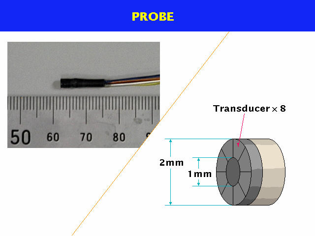
The following figure indicate the real and schematic view of the ring array probe. Each transducer can transmit and receive a ultrasonic spherical pulsed wave (resonance frequency 10MHz).
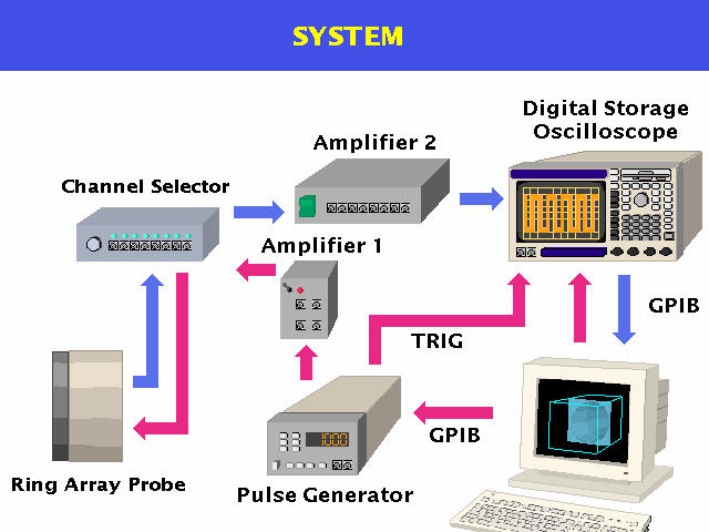
The following figure is a block diagram of the system which is used for the experiment shown in the next section.
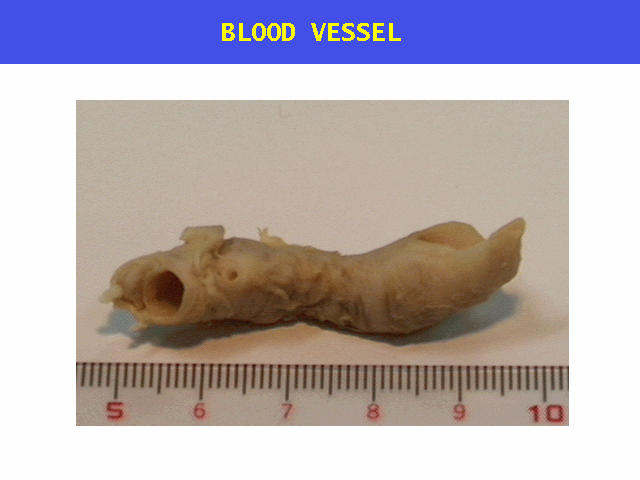
The blood vessel indicated in the following is a part of an artery of a human immersed in formalin. The middle part of the blood vessel was blockaded artificially.
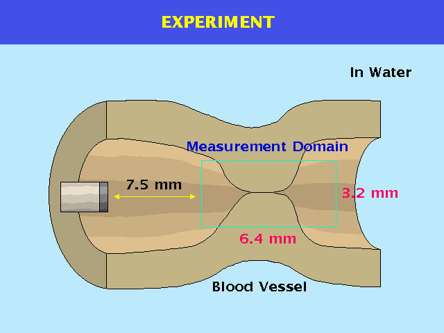
The blockaded part was visualized using our new method. The experiment was performed underwater. The measurement domain was indicated in the following figure. The inside of the measurement domain was quantized into 8*8*16 voxels.
The following figures are reconstructed images of the blood vessel. The reconstructed image has three-dimensional data, we can observe it from any viewpoint. An arrow indicates a direction of the probe and a wire frame indicates a measurement domain.
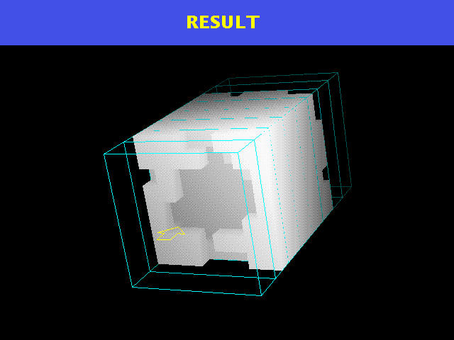
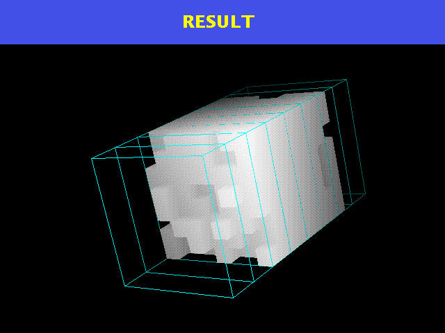
Beginning of the blockaded part is confirmed in front and an end part is confirmed backward. Using our new method, we can obtain frontal three-dimensional images at high speed. In theory, we can raise a frame rate up to about 4700 images/sec on our experimental condition. Therefore, our new method is suitable for observing objects which change the shape at high speed or for a probe which moves quickly. I hope that our new method will be applied to introvascular endoscopes in early stages.
 return to the home page
of this research
return to the home page
of this research